


|
Carnegie Mellon University |

|
Proximal Probe techniques development |
|
Proximal Probe Techniques (PPT) started almost two decades ago, with the introduction of Scanning Tunneling Microscopy (STM), followed few years later by Atomic Force Microscopy (AFM). Within this period they evolved from a tool of solid state physicists to a diverse group of tools showing especially great potential in the field of nanoscience of soft condensed matter. This potential is based on the current ability of PPTs to image organic (macro)molecules and their assemblies under a variety of conditions, especially in aqueous environments.
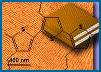
One of the major thrusts in proximal probe techniques is combination of imaging capabilities with simultaneous measurements of physical properties. In tapping mode atomic force microscopy (TMAFM), the most straightforward way to accomplish this goal is to reconstruct the time-resolved force interaction between the tip and surface. These tip-sample forces can be used to detect interactions (e.g., binding sites) and map material properties with nanoscale spatial resolution. Here, we describe a previously unreported approach, which we refer to as scanning probe acceleration microscopy (SPAM), in which the TMAFM cantilever acts as an accelerometer to extract tip-sample forces during imaging. This method utilizes the second derivative of the deflection signal to recover the tip acceleration trajectory. The challenge in such an approach is that with real, noisy data, the second derivative of the signal is strongly dominated by the noise. This problem is solved by taking advantage of the fact that most of the information about the deflection trajectory is contained in the higher harmonics, making it possible to filter the signal by "comb" filtering, i.e., by taking its Fourier transform and inverting it while selectively retaining only the intensities at integer harmonic frequencies. Such a comb filtering method works particularly well in fluid TMAFM because of the highly distorted character of the deflection signal. Numerical simulations and in situ TMAFM experiments on supported lipid bilayer patches on mica are reported to demonstrate the validity of this approach. Legleiter, J.; Park, M.; Cusick, B.; Kowalewski, T. PNAS, 2006, 103, 4813-4818
|
|
Room 527, 4400 Fifth Avenue Mellon Institute Pittsburgh, PA 15213 Phone: 412-268-9175 |