Refined Solution Structure and Ligand-binding Properties of PDC-109 Domain B. A Collagen-binding Type II Domain.
Constantine, K.L., Madrid, M., Banyai, L., Trexler, M., Patthy, L. & Llinás, M. J.Mol.Biol., 223, 281 (1992)
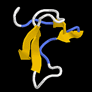
We have determined, via 1H-n.m.r., the solution conformation of the collagen-binding b-domain of the bovine seminal fluid protein PDC-109 (PDC-109/b). The structure determination is based on 341 interproton distance estimates and 42 dihedral angle estimates: a set of 24 initial structures were computed; 12 using the variable target function program DIANA, and 12 using the metric matrix program DISGEO. These structures were optimized by restrained energy minimization and dynamic simulated annealing using the CHARMM and X-PLOR programs. The average pairwise root-mean-square difference (r.m.s.d) between the optimized DIANA (DISGEO) structures is 0.71 Å (0.82 Å) for the backbone atoms, and 1.73 Å (2.03 Å) for all atoms. Both sets of structures exhibit the same global fold, secondary structure and placement of most non-polar side-chains. Two central antiparallel beta-sheets, which lie roughly perpendicular to each other, and two irregular loops support a large, partially exposed, hydrophobic surface that defines a putative binding site. A test of a hybrid relaxation matrix-based distance refinement protocol (MIDGE program) was performed using a normalized 250 millisecond NOESY spectrum. The resulting distances were input to the molecular mechanics/dynamics procedures mentioned above in order to optimize the DIANA structures. Our results indicate that relaxation matrix refinement of distances is most useful when used conservatively for identifying underestimated distance constraints. 1H-n.m.r. monitored ligand titration experiments revealed definite, albeit weak, binding interactions for phenethylamine and leucine analogs (Ka less than or equal to 25 M-1). Residues perturbed by ligand binding include Tyr7, Trp26, Tyr33, Asp34 and Trp39. These results suggest that PDC-109/b may recognize specific leucine and/or isoleucine-containing sequences within collagen. |